An ECM-Rich Capsule Limits Doxorubicin Entry into HCC Spheroids

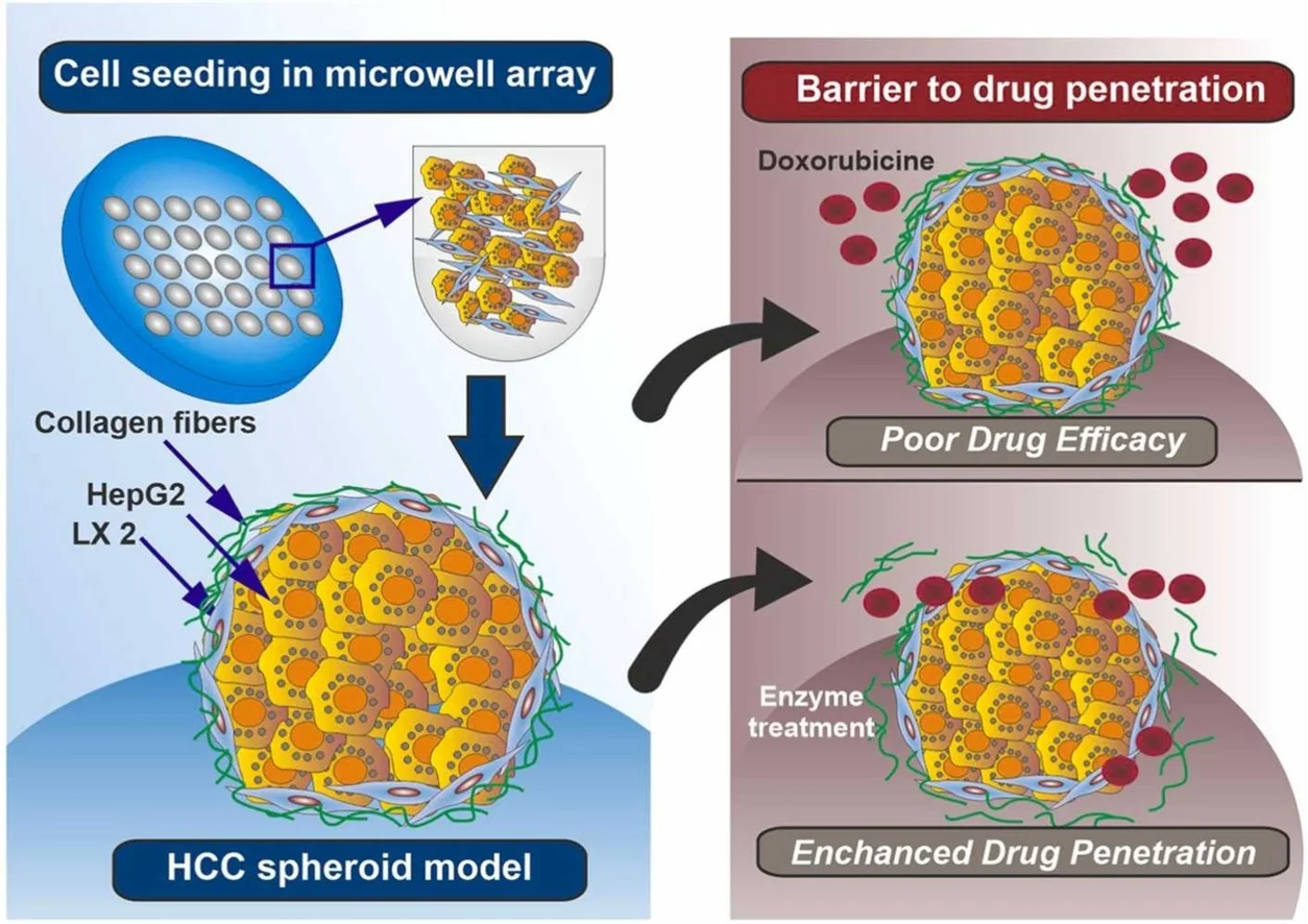
An academic team co-cultured HepG2 and hepatic stellate LX-2 cells to form a liver tumor spheroid model. Spheroids with a collagen-rich outer layer showed reduced doxorubicin penetration; disrupting that layer improved the drug response in the model.

Graphical abstract showing collagen-enclosed HepG2/LX2 spheroids and comparisons of drug penetration.
Graphical abstract of the collagen-capsule spheroid model and drug-penetration comparisons. (Żuchowska et al., CC BY 4.0)

Building a Spheroid with a Stromal Layer

Researchers at the University of Twente and collaborating medical centers used a soft-embossed microwell array to co-culture HepG2 hepatocellular carcinoma (HCC) cells with LX-2 hepatic stellate cells in different proportions. They cleared the resulting spheroids for imaging and stained for type I collagen to examine how cellular composition shaped the extracellular matrix (ECM).

Some compositions developed an extracellular-matrix-rich capsule around the spheroid. That feature matters to drug testing because the amount of drug reaching tumor cells may depend on the surrounding matrix, as well as on the cells’ intrinsic sensitivity.

Doxorubicin Meets the Matrix Barrier

The combined presence of hepatic stellate cells and extracellular matrix hindered doxorubicin penetration and reduced its therapeutic effect in vitro. Enzymatic degradation of the matrix-rich capsule improved penetration and the observed drug effect.

Sources

  1. Hepatocellular carcinoma (HCC) spheroid model exhibits a characteristic extracellular matrix-rich fibrous capsule hindering drug penetration. Biomedicine & Pharmacotherapy. 2026-10-21. DOI: 10.1016/j.biopha.2026.119998