Building the Models Took Time and Did Not Succeed for Every Sample
Researchers at the Luxembourg Institute of Health and collaborating hospitals used patient-derived three-dimensional spheroids for functional drug profiling in metastatic colorectal cancer. Spheroids were established from 12 of 19 patient samples, a 63% success rate. Two cultures were discarded after bacterial contamination; the others that failed could not be propagated. Among fresh samples used for screening, the median time from collection to release of drug results was 55.5 days, with two technical repeats extending the interval.
For one patient's model, responses to 33 drugs were compared before and after a freeze–thaw cycle. The dose–response area-under-the-curve values were strongly correlated (r = 0.9645). This is a matched comparison for one model, rather than evidence that every patient sample or repeated thawing cycle behaves the same way.
Checking What the Spheroids Retained
The investigators compared representative spheroids with the tumors from which they were derived before interpreting the drug screen. Histology and marker staining showed retained colorectal epithelial features, including CK20 and proliferative regions, although not every marker pattern was identical. These observations gave a tissue-level check that the growing culture still represented a relevant tumor population.
Genomic comparisons also found shared driver alterations and copy-number features, including changes in TP53, APC and KRAS and a MET amplification in the relevant sample. Some variant fractions were higher in spheroids, consistent with enrichment of cancer cells relative to the mixed original tissue. Conversely, some low-frequency variants in the source specimen were not detected in the model. The paired analyses therefore described both retained features and changes introduced by sampling or culture, rather than treating successful growth as proof that every tumor subclone had been preserved.
Drug Profiling Captured Some Tumor-Cell Differences
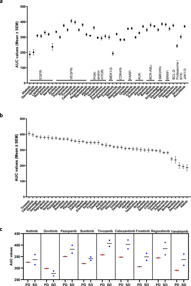
The wider screen included 42 anticancer small molecules and five standard-of-care agents. Spheroids carrying KRAS or BRAF alterations associated with resistance to EGFR-directed therapy generally showed low sensitivity to the relevant inhibitors. In the small comparison, anti-EGFR assay responses separated three cases classified as resistant by tumor mutations and one patient whose disease progressed on panitumumab from four patients whose disease was stable or regressive on anti-EGFR treatment. Clinical responses came from the last treatment before or first treatment after sample collection. Treatment combinations and timing differed between patients, so these data are exploratory rather than a calibrated prediction rule.
Eight spheroid samples were also tested for three days with 5-fluorouracil, SN-38, oxaliplatin and selected combinations. The two models from chemotherapy-naive tumors were relatively sensitive to standard-of-care treatments. Most other patients had previously received several lines of therapy, leaving the cohort weighted toward clinical nonresponders. Some ex vivo results agreed with the available treatment histories; others did not. The authors did not establish reliable predictive performance for these regimens.
The viability summaries used the area under a concentration–response curve. A high area meant that cells remained viable across the tested range, so it indicated relatively weak direct inhibition.
Antiangiogenic Therapy Fell Outside the Model's Strongest Use
Most VEGFR inhibitors produced little direct inhibition of spheroid viability. The clinical comparison for antiangiogenic treatment involved only three patients whose relevant regimen immediately preceded or followed sample collection: two had stable disease and one had progressive disease. Their treatment included chemotherapy plus bevacizumab, whereas the ex vivo assay measured small-molecule VEGFR inhibitors. The mismatch between clinical and ex vivo treatments, combined with the three-patient sample, limits what this comparison can show.
These tumor-only spheroids lacked a vascular compartment. A viability assay focused on cancer cells cannot capture a treatment's effect on blood-vessel formation in the surrounding tissue. The weak and discordant antiangiogenic readouts help define a limit of this particular model, without showing that antiangiogenic drugs lack clinical activity.