Fluorescence for Training, Bright-Field Images for Inference
Morphi is an image-analysis framework developed by researchers at Westlake University and packaged as a software tool. Using bright-field microscopy images, it estimates viability and maps regions of cell death within individual three-dimensional tumor models. The work was posted as a bioRxiv preprint on October 23, 2026, and has not been peer reviewed.
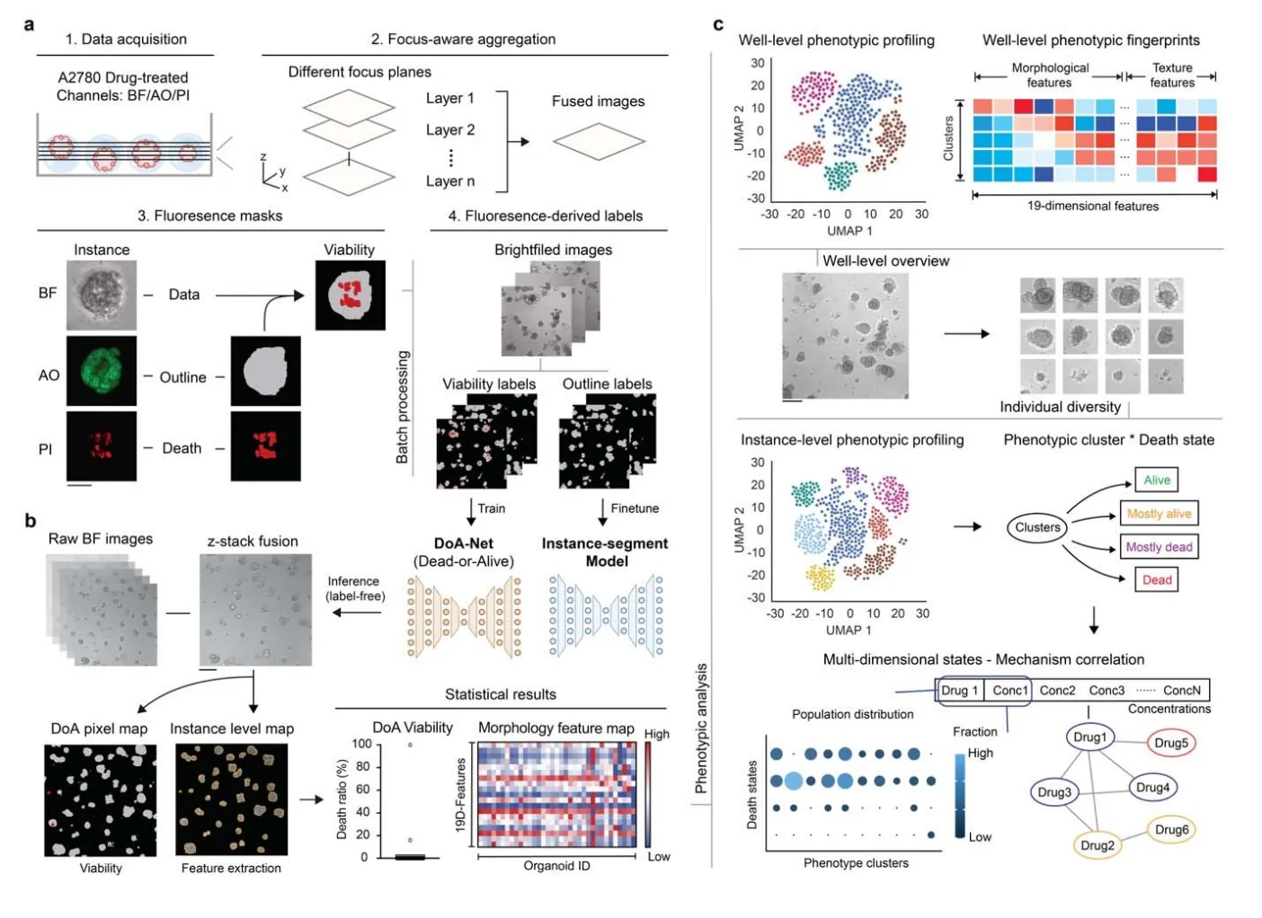
The training data came from three-dimensional spheroids grown from the A2780 ovarian cancer cell line. After treatment with 24 compounds for three days, the team collected matched bright-field images, fluorescence images to identify spheroid boundaries, and propidium iodide (PI) images to identify dead cells.
Fluorescence-derived masks supplied weak labels for training, avoiding dense manual annotation of live and dead regions. Morphi combines instance segmentation with DoANet, a neural network that assigns pixel-level viability estimates. Once trained, the model can perform inference from bright-field images alone.
Because each three-dimensional structure spans several focal planes, the workflow first combines the sharp regions of an image stack into a focus-enhanced projection. The initial dataset contained 1,510 multichannel stacks and more than 23,000 valid instances. After image-quality filtering, 6,897 instance images were split 80:20 for model development and testing, with 1,380 instances in the test set. Focus selection, object boundaries and debris removal are separate parts of the analysis.
Strong Overall Agreement, but Different Biological Readouts
Across the A2780 compound screen, the areas under drug-level bright-field dose–response curves correlated with fluorescence-based measurements at Pearson r = 0.95. Results from two independent dilution experiments correlated at r = 0.91, and agreement with the ATP-based CellTiter-Glo assay was r = 0.97.
Four drugs—AZD8055, temsirolimus, lapatinib and trametinib—produced different results in the two assays. CellTiter-Glo indicated substantial growth inhibition, while Morphi detected relatively little cell death. Image review showed smaller spheroids before, or without, pronounced death signals. For AZD8055, the response shifted from predominantly cytostatic at lower concentrations toward cytotoxic at higher concentrations.
Bright-field and fluorescence measurements also differed under some treatment conditions. Morphi estimated more cell death than PI staining for epirubicin, gemcitabine and etoposide. The authors observed severe fragmentation with weak PI signal in affected images and proposed that staining could underreport heavily damaged structures. These drug-specific differences occurred despite the high overall correlation between the measurements.
From Viability Scores to Phenotypic Fingerprints
The team tested transfer to WM0014, a gastric cancer patient-derived organoid model that was absent from training, and to an existing screen of 5,634 compounds across ten tumor organoid models. The analysis identified active compounds in those images, although the authors note that sensitivity and accuracy may differ between models.
Morphi also extracts 19 descriptors of size, shape, boundary structure and texture. At the well level, unsupervised analysis separated five states along a continuum from viable growth to cell death. One state consisted of smaller but largely viable, intact structures; others showed progressively fragmented boundaries and extensive death.
Dose-dependent trajectories differed between compounds. Paclitaxel produced an abrupt shift toward a disrupted state, whereas vinorelbine and gemcitabine passed through several intermediate states. These trajectories show how responses change with concentration, information that a single average viability value does not capture.
At the instance level, ten morphology clusters were combined with four predicted death states to form a 40-component representation of each well. Drug fingerprints sometimes aligned with known mechanisms and sometimes converged across different mechanisms, capturing functional similarities beyond conventional mechanism-of-action categories.