Review Weighs Image-Analysis Tools for Spheroid and Organoid Drug Screens

A review of bright-field and phase-contrast imaging finds different strengths and limits in rule-based, classical machine-learning and deep-learning analysis of spheroids and organoids. Dataset shifts, segmentation failures and two-dimensional projections remain practical concerns.

Black-and-white view of the University of Pretoria’s Hatfield campus.
The University of Pretoria’s Hatfield campus, photographed in 2024. (Cnrza UP, CC BY-SA 4.0)

No One Tool Class Fits Every Assay

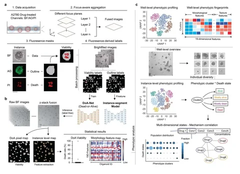
Three University of Pretoria researchers review automated analysis of bright-field and phase-contrast images from spheroids and organoids used in high-content screening. Rule-based approaches can standardize measurements that once required manual tracing, but may fail when imaging conditions change. Classical machine learning can handle some treatment-related changes in morphology, although it often needs retraining. Deep-learning tools learn from images more directly, yet their performance still depends on the training data and experimental setting.

Match the Analysis to the Measurement

The review recommends choosing tools according to the biological endpoint, image quality, culture shape, available annotations and computing resources. Most methods work from two-dimensional projections of three-dimensional structures, which can distort volume estimates for irregular spheroids or organoids. Performance also varies between data sets and imaging laboratories. The authors describe a choice of analytical approach tied to the assay and image set, rather than a universal ranking of software.

Sources

  1. Ncube KN, van den Bout I, Cordier W. Automated Spheroid and Organoid Image Analysis from Rule-Based Methods to Deep Learning. SLAS Discovery. 2026. DOI: 10.1016/j.slasd.2026.100344.