Conformable MEA Recording of DMD Cardiac Organoids
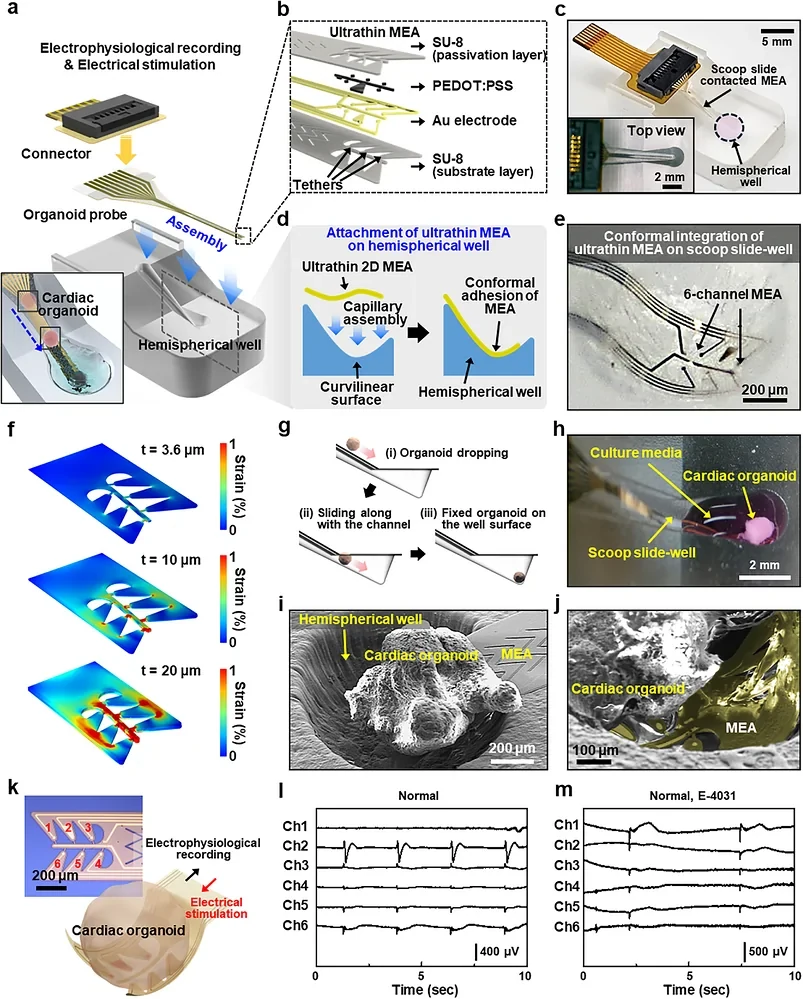
A flexible microelectrode array (MEA) and scoop-shaped well combine extracellular recording and video to compare cardiac organoids from healthy donors and patients with Duchenne muscular dystrophy (DMD).
Disease phenotyping and hERG-blocker response studies in patient-derived cardiac organoids.
Model & Design
Cardiac organoids derived from hiPSCs from two healthy donors and two DMD patients, containing cardiomyocytes and several non-cardiomyocyte populations.
Experimental Format
A scoop-shaped well supports the tissue on an approximately 4 μm-thick conformable array, with six-channel recording and simultaneous video.
Model Components
Component
Cardiac tissue
Description
Organoids formed without Matrigel or another exogenous extracellular matrix.
Component
Recording interface
Description
SU-8/Au/PEDOT:PSS flexible electrodes integrated with a PDMS well; 20 μm electrode diameter and 125 μm center-to-center spacing.
Component
Pharmacological and functional measurements
Description
E-4031 hERG blockade, extracellular field potentials, video-derived local motion and complementary calcium imaging.
Measurements & Endpoints
Extracellular field potentials
Field potential duration and rhythm
Regional tissue motion
Calcium activity
Cell composition and disease-associated markers
Analysis
Identify electrical features after denoising and compare them with video-derived motion. Calculate field potential duration only when repolarization features are identifiable; use single-cell data to characterize donor differences.
Published Evidence
Study Design
Structural and functional comparison of healthy-donor and DMD organoids, including responses to E-4031.
Study Sample
Four donors; more than 40 organoids were pooled per donor sample for single-cell analysis.
Comparison
Healthy-donor organoids, pretreatment baseline and drug-exposure conditions.
Finding
DMD organoids showed extracellular-matrix and cellular-composition changes and greater vulnerability to drug-associated rhythm instability.
Interpretation & Limits
The study examined four donors and one probe drug; cardiac tissue retained immature features.
Afterdepolarization-like extracellular signals are distinct from intracellular action-potential measurements. Video measured motion rather than force.
Manual loading and irregular DMD waveforms limit throughput and automated interval analysis.
A platform built around a flexible microelectrode array and scoop-shaped well combines electrical recordings with video of tissue motion. Organoids from healthy donors and patients with Duchenne muscular dystrophy show different structural features and responses to hERG blockade.