Improving Contact with a Three-Dimensional Tissue
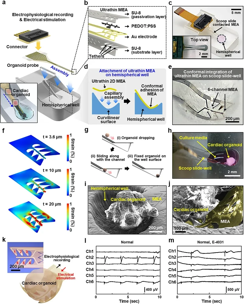
A curved organoid surface does not make uniform contact with a flat electrode array. Researchers at Pusan National University and collaborating institutions addressed that mismatch with an ultrathin microelectrode array (MEA) integrated into a scoop-shaped well. In Advanced Science, the team describes using the device to record electrical activity in cardiac organoids while observing their motion.
The well guides an organoid toward the recording region under gravity, while the flexible electrode layer conforms to the curved surface beneath it. The approximately 4 μm-thick device has six recording channels, with electrodes 20 μm in diameter. It records extracellular field potentials while permitting optical observation and changes in drug exposure.
The study applied this interface to organoids derived from healthy donors and people with Duchenne muscular dystrophy (DMD), a condition marked by dystrophin deficiency that can severely affect the heart. The aim was to compare spontaneous electrical activity, movement and responses to pharmacological stress in the same tissue.
Four Donors, with Different Cellular and Structural Features
The organoids came from induced pluripotent stem cells from two healthy donors and two DMD patients. The study confirmed pathogenic DMD variants and characterized the donor lines. Organoid formation did not use Matrigel or another exogenous extracellular matrix.
Single-cell RNA sequencing pooled more than 40 organoids per donor sample and identified eight major cell populations, including cardiomyocytes, fibroblast populations and endothelial-related cells. Further analysis separated cardiomyocyte subtypes. Both overall cellular composition and subtype proportions varied between donors.
DMD organoids lacked dystrophin and showed changes in collagen- and extracellular-matrix-associated expression. Vascular features and the spatial distribution of cardiomyocytes also differed: healthy tissues had a more continuous arrangement, whereas DMD tissues included localized clusters and fragmented organization.
Both groups retained features of developing cardiac tissue, including low levels of ventricular maturation markers. These structural and developmental features are relevant to interpreting electrical differences alongside the disease genotype.
Electrical Activity and Motion Recorded Together
The array captured extracellular signals on the millivolt scale. After denoising, the team identified depolarization- and repolarization-associated features and calculated field potential duration where the waveform supported it. Multiple channels sampled different regions of the organoid.
Video analysis tracked changes in pixel intensity within selected regions to describe tissue motion over time. Combining these records with the electrical traces allowed the researchers to compare electrical events with contraction and relaxation patterns. The optical readout measured movement rather than contractile force.
Additional calcium imaging showed faster-rising, higher-amplitude calcium signals in DMD organoids. Single-cell data revealed altered ion-channel expression and cardiomyocyte subtype composition, providing context for the irregular electrical behavior.
Different Responses to hERG Blockade
Pharmacological testing used E-4031, a blocker of the hERG potassium channel. In analyzable healthy-donor recordings, exposure prolonged repolarization-related field potential measures. DMD organoids showed greater vulnerability to electrical instability and irregular rhythm during treatment.
Some DMD traces contained additional deflections after the repolarization phase, which the authors describe as afterdepolarization-like activity. Once a distinct repolarization feature was lost, interval measurements became unreliable. Raw waveform shape, rhythm and motion records therefore remained important alongside calculated field potential duration.
The two patients had different clinical cardiac presentations; one had a history of ventricular hypertrophy. The organoid comparisons explore how these donor-associated differences appear under the same pharmacological challenge.
Manual Loading Limits Throughput
The platform combines organoid positioning, conformable electrodes and optical imaging. It permits repeated observation of the same three-dimensional cardiac organoid’s electrical and mechanical behavior under changing conditions.
Manual loading and alignment remain a bottleneck and can introduce operator variation. The authors propose automated positioning, improved contact across tissue shapes and algorithms that can handle irregular DMD waveforms. Additional calcium or force measurements could further clarify the relationship between electrical activity and tissue mechanics.