Electrical and Contraction Assessment in an ET-1 hiPSC-Cardiomyocyte Model
Endothelin-1 (ET-1)-induced remodeling is assessed by gene expression, patch clamp and video-motion analysis to test selective responses to Cav1.3 C-terminal delivery.
Compare intervention effects on repolarization, ion currents and motion-derived contraction in a human heart-failure-like cell model.
Model & Design
Cardiomyocyte monolayers derived from one male and one female human induced pluripotent stem cell (iPSC) line.
Experimental Format
ET-1 exposure at 10 nM for 48 hours from day 20; the intervention group receives AAV6-Cav1.3 C-terminal delivery for 48 hours from day 22. Untreated, ET-1 and intervention groups are assessed on day 30.
Model Components
Component
Human cardiomyocytes and remodeling stimulus
Description
Cardiomyocytes derived from two hiPSC lines; ET-1 induces molecular, electrical and contraction changes.
Component
Gene-delivery component
Description
AAV6 carries the human Cav1.3 C-terminal fragment spanning amino acids 1840–2137.
Component
Functional measurements
Description
RT-qPCR, single-cell patch clamp and phase-contrast particle tracking; video supplies contraction-amplitude measurements.
Measurements & Endpoints
Hypertrophy and ion-channel gene expression
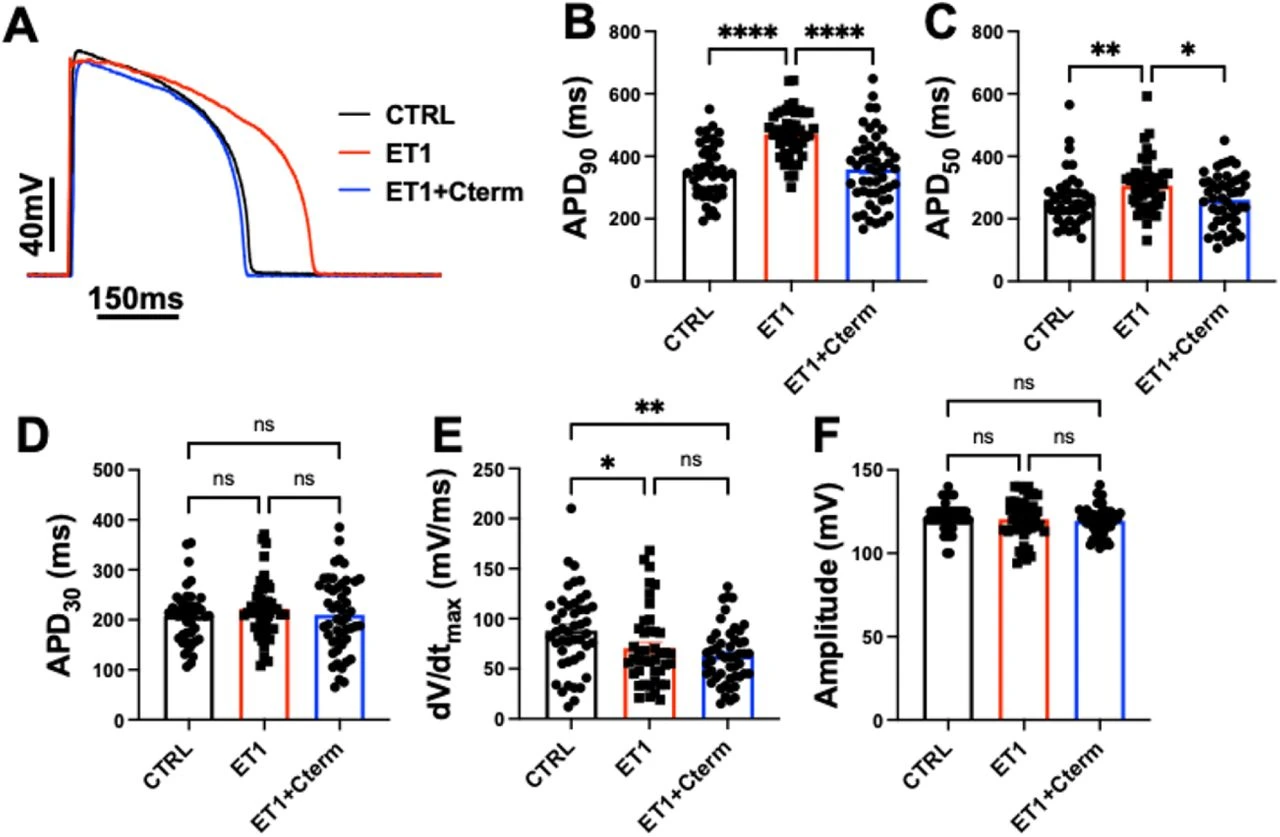
Action-potential repolarization and upstroke velocity
Outward potassium, L-type calcium and sodium-current densities
Membrane capacitance
Spontaneous beating, video-derived contraction amplitude and kinetics
Analysis
Evaluate molecular, single-cell electrical and monolayer-motion endpoints separately, retaining persistent sodium-current, upstroke and hypertrophy-related changes.
Published Evidence
Study Design
Parallel three-group comparison of ET-1 remodeling and the response to C-terminal delivery.
Study Sample
Two cell lines; action-potential recording n = 42/46/50 for untreated, ET-1 and intervention groups. Electrophysiology/contraction captions specify three independent differentiations; the expression figure separately specifies four biological replicates.
Comparison
Untreated cells, ET-1 alone and ET-1 plus the C-terminal fragment.
Finding
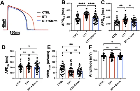
ET-1 prolonged APD90/APD50 by 34%/17%. Fragment delivery restored repolarization, measured potassium/calcium currents and video contraction amplitude; sodium-current, upstroke and capacitance abnormalities persisted.
Interpretation & Limits
Preprint; two cell lines, with differentiation replicates distinguished from recording counts.
Immature hiPSC-CM features and limited monoculture complexity remain.
Video measures motion-derived amplitude; direct versus indirect KCNH2 regulation is unresolved.
A preprint reports recovery of selected calcium and potassium currents, repolarization and video-derived contraction after delivery of a Cav1.3 fragment. Sodium-current and hypertrophy-related changes persisted.