A Human-Cell Model Tests a Specific Variant
A study in Human Mutation used induced pluripotent stem cell-derived cardiomyocytes from one person carrying the CAV3 p.Ala46Thr variant. The donor had skeletal-muscle symptoms without a clear cardiac abnormality at the recorded clinical assessment. The comparison cells came from an unaffected sister who did not carry the variant. These are related donors, but the cells are not an isogenic gene-edited pair.
The researchers checked the induced cell line's genotype and pluripotency before differentiating it into cardiomyocytes. They then compared cellular structure, CAV3 protein localization and calcium activity before and after the beta-adrenergic agonist fenoterol. CAV3 encodes caveolin-3, a membrane-associated protein that helps organize caveolae and signaling complexes in muscle cells.
Protein Localization Diverged More Clearly Than Basic Structure
Cardiomyocyte markers and electron microscopy showed broadly recognizable cardiac-cell structures in both groups, including organized myofibrils, Z-lines and mitochondria. Sarcomere length and cell area did not differ significantly, although cell perimeter did. Visible membrane caveolae remained in the patient-derived cells, and the density of these indentations was not significantly different between groups.
CAV3 localization differed more sharply. The protein was seen at the membrane and in the cytoplasm of control cells, but accumulated in or around the nucleus of patient-derived cells, with little membrane signal. Abnormal localization was recorded in 20 of 23 patient-derived cells and none of 22 control cells examined.
Faster Calcium Transients Did Not Restore Rhythm Regularity
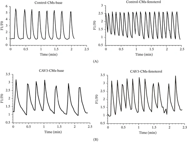
Using Fluo-4 calcium imaging, the team measured slower and less regular baseline activity in patient-derived cardiomyocytes. In the reported recordings, control cells rose from 4.0 to 7.0 transients per minute after fenoterol, while patient-derived cells rose from 2.8 to 4.4. The coefficient of variation of the beating intervals was 1.87% before and 0.97% after fenoterol in control cells, versus 22.73% and 31.42% in patient-derived cells. Frequency increased in both groups, but regularity did not improve in the variant-bearing model.
Calcium imaging included 13 cardiomyocytes per group from at least three independent experiments. The authors lacked systematic serial electrocardiograms and echocardiograms for the family, preventing alignment of the cellular irregularity with a measured course of cardiac disease. One donor pair and a non-isogenic control also left genetic-background effects and the variant’s contribution intertwined. The result is a specific human-cell drug-response comparison rather than an isolated causal test or a forecast of clinical cardiac outcomes.