Generating Two Models from the Same Tumor
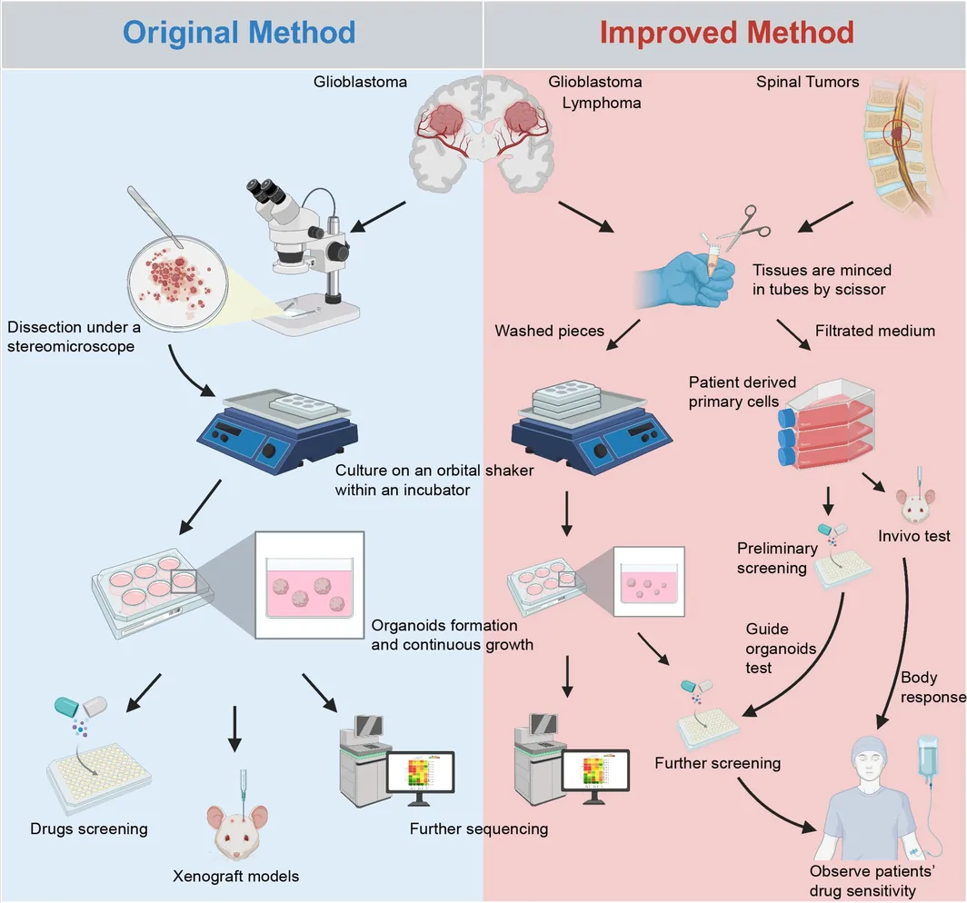
Researchers at Renji Hospital, Shanghai Jiao Tong University School of Medicine, and collaborating institutions reported an adapted workflow for patient-derived glioblastoma models in CNS Neuroscience & Therapeutics. The approach aims to simplify the preparation of three-dimensional organoids while recovering matched primary tumor cells.
The study collected 22 central nervous system tumor specimens, including 18 glioblastomas. Organoids were established from 15 of the 18 glioblastoma specimens, approximately 83%. Most formed within one to two weeks, and the report describes subsequent passage, storage and recovery.
Primary cells were obtained from material collected during organoid preparation. Retaining this material provided a paired cellular model from the same patient, alongside organoids suited to examining tissue structure and heterogeneous responses. The two formats offer complementary material for drug and mechanistic studies.
Comparing Tissue Features and Genomic Variants
Histological assessment showed preservation of some features of the parental tumor, including nuclear atypia. Related features remained visible after passage and recovery from storage.
In the representative matched whole-exome comparison, organoids shared 86.8% of single-nucleotide variants with the parental tumor. Overlap between original organoids and passaged or recovered material was 88.8% and 88.4%, respectively.
The primary cells were evaluated by morphology and CD133 expression. The authors reported a higher CD133-positive proportion than with the traditional isolation approach used as a comparator.
Temozolomide Responses Measured as Projected Area
The drug experiment compared 160 μM temozolomide with a vehicle control in organoids from two patients, using three organoid replicates per treatment. According to the Methods, exposure lasted one week, followed by withdrawal and a further two weeks of observation.
The readout was the two-dimensional projected area of each organoid in bright-field images and its change over time. The two patient-derived models showed different degrees of growth response.
Clinical follow-up was available for only one of the two patients; that patient experienced recurrence after one year of temozolomide treatment. The other was lost to follow-up. The experiment therefore provides a small exploratory comparison rather than an estimate of clinical predictive accuracy.
Initial Extension to Other Central Nervous System Tumors
The team also reported three-dimensional models from subependymoma and diffuse large B-cell lymphoma specimens. Their appearance, growth and culture requirements differed from glioblastoma organoids. In the discussion, the authors note that lymphoma structures could persist in glioblastoma organoid medium without sustained growth.
These observations suggest that forming a three-dimensional structure does not guarantee a usable long-term model. Culture requirements would need to be adapted to the tumor type before broader drug-response comparisons.
Recurrent Samples and Vascular Features Remain Challenges
The authors found it more difficult to establish organoids from recurrent glioblastoma after chemoradiotherapy. The current tissues also had limited vascular components, which can influence tissue organization and drug access.
Paired primary cells extend the available experimental material, but prolonged adherent culture can alter stem-like features. The approach preserves related two- and three-dimensional patient-derived models, while its use in drug testing remains exploratory.