From Organoid Cell Loss to Intestinal Injury
An AstraZeneca team developed a framework combining human small-intestinal organoids with mathematical models to investigate diarrhea associated with oral oncology drugs. Published in CPT: Pharmacometrics & Systems Pharmacology, the study focuses on epithelial injury caused by antiproliferative activity.
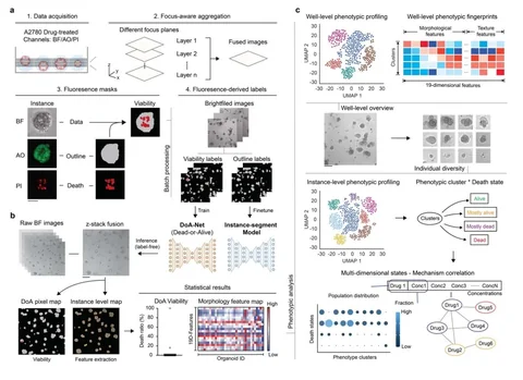
The six drugs were the CDK4/6 inhibitors abemaciclib and ribociclib and the EGFR inhibitors poziotinib, lazertinib, mobocertinib and osimertinib. Organoids came from adult intestinal stem cells from one donor. Imaging at multiple concentrations and time points distinguished proliferative from non-proliferative cell populations.
A mathematical model described organoid growth, transitions to non-proliferative states, cell death and eventual culture decline. The researchers incorporated drug effects on cell-cycle arrest and death into this model, then transferred those effects to a model of human intestinal epithelial turnover. This separates the measured cellular response from the physiological process that turns it into tissue injury.
Different Responses Within Drug Classes
Abemaciclib markedly reduced both proliferative and non-proliferative cell counts. Ribociclib produced only a small reduction at the highest tested concentration. The contrast was consistent with the higher clinical diarrhea burden reported for abemaciclib and provided a test of whether the framework could distinguish drugs in the same class.
The four EGFR inhibitors also affected both cell populations to varying degrees. The investigators modeled these changes as drug-induced death of proliferative cells. This is a model of antiproliferative epithelial injury; gastrointestinal effects driven by other mechanisms would require corresponding mechanistic information.
Plasma and Intestinal Exposure Predict Different Levels of Injury
The human epithelial model follows stem and proliferative progenitor cells in the crypt and non-proliferative cells in the crypt and villus. It describes tissue renewal, cell loss and recovery after a drug challenge. Physiologically based pharmacokinetic (PBPK) modeling supplies the exposure profile used to drive that response.
The concentration of free drug in human crypt cells is difficult to measure directly. The team therefore compared two surrogates: unbound plasma concentration and unbound enterocyte concentration. During oral absorption, local intestinal exposure can exceed plasma exposure. The enterocyte scenario used a multilayer gut-wall model within the PBPK platform to represent that possibility.
For abemaciclib, Table 2 reports predicted villus injury of 1.8–2.6% with plasma exposure and 46.5–51.8% with enterocyte exposure across the tested regimens. For mobocertinib, the corresponding estimates were 7.2% and 100%. For poziotinib and osimertinib, both exposure scenarios reached concentrations associated with injury. Ribociclib and lazertinib produced relatively small modeled effects under either scenario.
The exposure model estimates drug concentrations near the cells; the turnover model estimates the resulting changes in proliferation, differentiation and cell loss. Together, they track injury and recovery over a dosing regimen.
Relating Injury to Clinical Diarrhea
The investigators expressed villus injury as the proportion of missing epithelial cells and related it to clinical reports of any-grade and grade 3 or higher diarrhea. A nonlinear statistical model was fitted to the study’s clinical data to translate the predicted injury into a probability of diarrhea.
For each regimen, the maximum predicted probability during treatment was compared with the reported clinical incidence. Enterocyte-based exposure gave closer agreement across the six drugs than plasma exposure, which failed to predict substantial intestinal injury for two drugs with important clinical diarrhea findings.
The injury-to-diarrhea relationship was fitted using this same set of drugs and clinical observations. Its performance on additional drugs remains to be tested independently. Likewise, neither exposure surrogate was a direct measurement of free drug in the crypt.
The authors discuss other contributors to EGFR-inhibitor diarrhea, including secretory pathways, as well as variation between patients and clinical trials. Extending the framework will require a broader range of compounds and population-level descriptions of exposure, epithelial injury and recovery. The current study demonstrates the importance of local exposure when interpreting human in vitro toxicity for orally administered drugs.